


Study: ‘Alleged Virus’ Is Identical to Normal Cell ‘Structures’
It's ALL FRAUD!!
Dear friends,
Thanks to the diligent efforts of one of my listeners, I received a paper yesterday that puts another nail in the coffin for the existence of SARS-CoV-2. The paper is titled “Appearances Can be Deceiving – Viral-like Inclusions in Covid-19 Negative Renal Biopsies by Electron Microscopy.” The authors are Clarrisa A. Cassol, et al., and the citation is Kidney360 1:824-828, 2020. This is a peer-reviewed journal affiliated with the American Society of Nephrology; in other words, this paper comes squarely from what is called acceptable, mainstream science.
Many of you have probably seen the electron-micrograph pictures of SARS-CoV-2, the ones in black and white, with the black dots within the faint outline of the circle. I have attached two such images from papers that claim these photos show direct evidence of the existence of the virus. These are the pictures that virologists show us, not the computer-generated, colorful images that you see in magazines and on the internet. These are the “real” pictures of the virus, and they are offered as “proof” that the virus exists.
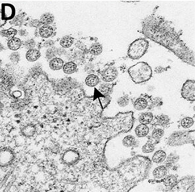
However, it turns out these photos are actually NOT corona viruses, and the CDC, among others, has known this fact since at least 2004. The above paper examines the evidence used to claim that these images represent viruses, rather than normal “structures” within a cell, particularly sick cells. Here is what the paper says:
“We have observed morphologically indistinguishable inclusions within podocytes and tubular epithelial cells both in patients negative for coronavirus disease 2019 (COVID-19) as well as in renal biopsies from the pre-COVID-19 era” (emphasis added).
In other words, the researchers saw these same structures in people with no evidence of Covid and in samples they took before Covid even happened, before the virus was said to even exist.
In addition, they say:
“We postulated that endogenous mimickers could be present that are morphologically indistinguishable from SARS-CoV-2 virions ultrastructurally.”
And:
“Viral-like inclusions, consisting both of single vesicles with diameters between 50 and 138 nm, as well as packed groups within larger vesicles, were found in all 15 cases, either in podocytes. Tubular epithelia, or vascular epithelial cells (figure 1).”
In all 15 cases that they examined, they found structures identical to what is being called SARS-CoV-2. They were scattered all over the kidneys and blood vessels; they are not viruses, but normal parts of the cells.
Then they go on to describe how these particles come about:
“A number of potential natural mimickers that can generate intracellular groups of round vesicles mimicking
SARS-CoV-2 virions could be listed, the most likely being endocytic vesicles and endosomal components such as microvesicular bodies containing exosomes, among others. Endocytosis leads to the formation of 60-120 nm vesicles, which is within the size range described for SARS-CoV-2 (60-140nm). These endocytic vesicles may be coated by different proteins, one of the most common being clathrin. The presence of coating proteins may be responsible for the presence of an electron-dense area surrounding these vesicles, giving the appearance of a viral corona.”
In other words, remember the famous “corona” on the corona virus? It turns out it’s just a common protein coating on normal vesicles, picking up the dyes in the electron-microscope preparation. The corona appearance is just another creative fiction, dreamed up by virologists and their graphic design teams.
Finally, the paper goes on to say that, naturally, you see more of these particles in sick people than in healthy people, which is exactly what I have been suggesting this past year. Dead and dying cells make these particles in the dying process and partly to get rid of poisons.
But the final nail comes in this quote:
“The potential for confusion of coronavirus particles with normal cellular components was in fact highlighted in a detailed ultrastructural study by the Centers for Disease Control and Prevention (CDC) of SARS-CoV responsible for the 2003 SARS outbreak.”[1]
In other words, the CDC in 2004 knew that researchers couldn’t reliably know these particles were coronavirus particles. Not a word has been heard about this since. All virologists use these pictures as proof of the existence of this virus. It is a fraud, based on junk science, like everything else connected with “Covid 19.”
[1] GoldsmithCS, Tatti, TD, Ksiazek TG, Rollin PE, Comer JA, Lee WW, Rota PA, Bankamp B, Belini WJ, Saki, SR: Ultrastructural characterization of SARS coronavirus. Emerg Infect Dis 10: 320-326, 2004.